| dc.contributor.author | Tunçer, Özlem Güngör | |
| dc.contributor.author | Bekmezci, Yavuz | |
| dc.contributor.author | Karadereler, Selhan | |
| dc.contributor.author | Karakoç, Çağla | |
| dc.contributor.author | Altındağ, Ebru | |
| dc.contributor.author | Tolun, Reha | |
| dc.contributor.author | Krespi, Yakup | |
| dc.date.accessioned | 2016-12-12T12:24:10Z | |
| dc.date.available | 2016-12-12T12:24:10Z | |
| dc.date.issued | 2012 | |
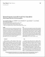
| dc.identifier.citation | Tuncer OG, Yavuz Bekmezci, Karadereler S, Karakoc C, Altındag E, Tolun R, Krespi Y. Epidural empyema caused by frontal sinus aspergillosis. Archives of Neuropsychiatry 2012; 49(1): 83-5. doi: 10.4274/npa.y6167 | en_US |
| dc.identifier.issn | 1300-0667 | |
| dc.identifier.uri | http://www.noropsikiyatriarsivi.com/sayilar/411/buyuk/83-85.pdf | en_US |
| dc.identifier.uri | https://hdl.handle.net/11446/1203 | en_US |
| dc.description | İstanbul Bilim Üniversitesi, Tıp Fakültesi. | en_US |
| dc.description.abstract | Aspergillosis infection located primarily in the frontal sinus and invading into the
dura is rare. A 74-year-old male patient was admitted with headache of 3-month
duration that did not respond to analgesics. Cranial MRI showed an irregular
hyperintense lesion in T2 FLAIR and T1 FLAIR, arising from the right frontal sinus
and advancing to the intracranial space. A collection, hyperintense on T2 FLAIR
and isointense on T1 FLAIR, adjacent to the left frontal sinus was observed,
consistent with epidural empyema. The lesions were drained surgically, and
subsequently treated with antifungal therapy using Amphotericin B. The headache
resolved completely after surgical drainage. The patient has been followed-up for
a year and is currently asymptomatic. Aspergillosis infections lodging primarily in
the frontal sinus and spreading to the dura are very rare, therefore, we report this
case in the present paper. ( | en_US |
| dc.description.abstract | Primer olarak frontal sinüse yerleşerek duraya yayılım gösteren aspergillosis infeksiyonu oldukça nadirdir. Yetmiş dört yaşında erkek hasta 3 aydır olan analjezik tedaviye cevap vermeyen baş ağrısı yakınmasıyla başvurdu. Kranyal MR’ında; sağ frontal sinüsten intrakranyal alana doğru uzanan T2 FLAIR ve T1 FLAIR‘de hiperintens irregüler lezyon olduğu, intraserebral alana doğru ilerlediği görüldü. Sol frontal bölgede ise T2 FLAIR’de hiperintens, T1 FLAIR’de ve GRE (ECHO) incelemesinde izointens görünen epidural ampiyem ile uyumlu kolleksiyon saptandı. Cerrahi olarak lezyonlar drene edildi, amfoterisin B tedavisi başlandı. Cerrahi drenaj sonrası bir yıllık takibi boyunca benzer başağrısı tekrarlamadı. Primer olarak frontal sinüse yerleşerek duraya doğru yayılım gösteren ve epidural ampiyeme neden olan aspergillosis infeksiyonlarının çok nadir olması nedeniyle bu olgu sunulmaktadır. | en_US |
| dc.language.iso | eng | en_US |
| dc.publisher | Aves Yayıncılık | en_US |
| dc.identifier.doi | 10.4274/npa.y6167 | en_US |
| dc.rights | info:eu-repo/semantics/openAccess | en_US |
| dc.subject | frontal | en_US |
| dc.subject | sinus | en_US |
| dc.subject | epidural | en_US |
| dc.subject | aspergillosis | en_US |
| dc.subject | empyema | en_US |
| dc.subject | ampiyem | en_US |
| dc.title | Epidural empyema caused by frontal sinus aspergillosis | en_US |
| dc.title.alternative | Epidural ampiyeme neden olan frontal sinüs aspergillomu | en_US |
| dc.type | article | en_US |
| dc.relation.journal | Nöropsikiyatri Arşivi / Archives of Neuropsychiatry | en_US |
| dc.department | DBÜ, Tıp Fakültesi | en_US |
| dc.identifier.issue | 1 | |
| dc.identifier.volume | 49 | |
| dc.identifier.startpage | 83 | |
| dc.identifier.endpage | 85 | |
| dc.contributor.authorID | TR118242 | en_US |
| dc.contributor.authorID | TR141267 | en_US |
| dc.contributor.authorID | TR223765 | en_US |
| dc.contributor.authorID | TR143782 | en_US |
| dc.contributor.authorID | TR140963 | en_US |
| dc.relation.publicationcategory | Belirsiz | en_US |